The NMR spectroscopy method is based on the magnetic properties of nuclei. The nuclei of atoms carry a positive charge and rotate around their axis. The rotation of the charge leads to the appearance of a magnetic dipole.
The angular momentum of rotation, which can be described by the spin quantum number (I). The numerical value of the spin quantum number is equal to the sum of the spin quantum numbers of protons and neutrons that make up the nucleus.
The spin quantum number can take on the value
If the number of nucleons is even, then I = 0, or an integer. These are such nuclei C 12 , H 2 , N 14 , such nuclei do not absorb radio frequency radiation and do not give signals in NMR spectroscopy.
I = ± 1/2 H 1 , P 31 , F 19 - absorb radio frequency radiation, give an NMR spectrum signal.
I = ± 1 1 / 2 CL 35 , Br 79 - asymmetric distribution of charges over the surface of the nucleus. This results in a quadrupole moment. Such nuclei are not studied by NMR spectroscopy.
PMR - spectroscopy
The numerical value of I (I = ± 1/2) determines the number of possible orientations of the nucleus in an external magnetic field in accordance with the formula:
This formula shows that the number of orientations is 2.
In order to carry out the transition of a proton located at a lower level to a higher one, it needs to be given an energy equal to the difference in the energy of these levels, that is, to be irradiated with radiation of a strictly defined purity. The difference in energy levels (ΔΕ) depends on the magnitude of the applied magnetic field (H 0) and the magnetic nature of the nuclei, which is described by the magnetic moment (μ). This value is determined by rotation:
![]() , where
, where
h is Planck's constant
The magnitude of the external magnetic field
γ is the coefficient of proportionality, called the gyromagnetic ratio, determines the relationship between the spin quantum number I and the magnetic moment μ.
![]()
– basic NMR equation, it relates the magnitude of the external magnetic field, the magnetic nature of the nuclei and the purity of the radiation at which the absorption of radiation energy occurs and the nuclei pass between levels.
From the above entry it can be seen that for the same nuclei, protons, there is a strict relationship between the value of H 0 and μ.
So, for example, in order for the proton nuclei to be in an external magnetic field of 14000 gauss, they need to be irradiated with a frequency of 60 MHz, if up to 23000 gauss, then radiation with a frequency of 100 MHz will be required.
Thus, from the above it follows that the main parts of the NMR spectrometer should be a powerful magnet and a source of radio frequency radiation.
The analyte is placed in an ampoule made of special grades of glass 5 mm thick. The ampoule is placed in the gap of the magnet, for a more uniform distribution of the magnetic field inside the ampoule, it rotates around its axis, with the help of a coil, the radiation is generated by radio frequency radiation continuously. The frequency of this radiation varies in a small range. At some point in time, when the frequency exactly corresponds to the equation of NMR spectroscopy, absorption of radiation energy is observed and the protons reorient their spin - this absorption of energy is recorded by the receiving coil in the form of a narrow peak.
In some models of the spectrometer, μ=const, and in small aisles, the value of H 0 changes. To register the spectrum, 0.4 ml of a substance is needed, if the substance is a solid, it is dissolved in a suitable solution, 10-50 ml/g of the substance must be taken.
To obtain a high-quality spectrum, it is necessary to use solutions with a concentration of 10 - 20%. The NMR sensitivity limit corresponds to 5%.
To increase the sensitivity using a computer, many hours of signal accumulation are used, while the useful signal increases in intensity.
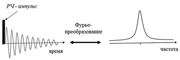
With the further improvement of the technique of NMR spectrum distribution, the use of Fourier - signal transformation has become. In this case, the sample is irradiated not with radiation with a slowly changing frequency, but with radiation that combines all frequencies in one packet. In this case, radiation of one frequency is absorbed, and the protons go to the upper energy level, then a short pulse is turned off and after that the excited protons begin to lose the absorbed energy and go to the lower level. This energy phenomenon is registered by the system in the form of a series of millisecond pulses that decay in time.
The ideal solvent is a substance that does not contain protons, that is, four carbon chloride and sulfuric carbon, however, some substances do not dissolve in these solutions, therefore, any solvents in the molecules of which the atoms of the light isotope H 1 are replaced by atoms of the heavy deuterium isotope are used. The isotopic frequency should correspond to 99%.
СDCl 3 - deuterium
Deuterium in the NMR spectra does not give a signal. A further development of the method was the use of a high-speed computer and signal fury conversion. In this case, instead of the last scan of the radiation frequency, instantaneous radiation containing all possible frequencies is superimposed on the sample. In this case, instantaneous excitation of all nuclei and reorientation of their spins occur. After turning off the radiation, the nuclei begin to emit energy and go to a lower energy level. This burst of energy lasts for several seconds and consists of a series of microsecond pulses, which are registered by the registration system in the form of a fork.
1. The essence of the phenomenon
First of all, it should be noted that although the word “nuclear” is present in the name of this phenomenon, NMR has nothing to do with nuclear physics and has nothing to do with radioactivity. If we talk about a strict description, then one cannot do without the laws of quantum mechanics. According to these laws, the interaction energy of a magnetic core with an external magnetic field can take only a few discrete values. If magnetic nuclei are irradiated with an alternating magnetic field, the frequency of which corresponds to the difference between these discrete energy levels, expressed in frequency units, then the magnetic nuclei begin to move from one level to another, while absorbing the energy of the alternating field. This is the phenomenon of magnetic resonance. This explanation is formally correct, but not very clear. There is another explanation, without quantum mechanics. The magnetic core can be thought of as an electrically charged ball rotating around its axis (although, strictly speaking, this is not the case). According to the laws of electrodynamics, the rotation of a charge leads to the appearance of a magnetic field, i.e., the magnetic moment of the nucleus, which is directed along the axis of rotation. If this magnetic moment is placed in a constant external field, then the vector of this moment begins to precess, i.e., rotate around the direction of the external field. In the same way, the spinning wheel axis precesses (rotates) around the vertical, if it is unwound not strictly vertically, but at a certain angle. In this case, the role of the magnetic field is played by the gravitational force.
The precession frequency is determined both by the properties of the nucleus and by the strength of the magnetic field: the stronger the field, the higher the frequency. Then, if, in addition to a constant external magnetic field, an alternating magnetic field acts on the nucleus, then the nucleus begins to interact with this field - it, as it were, swings the nucleus more strongly, the precession amplitude increases, and the nucleus absorbs the energy of the alternating field. However, this will occur only under the condition of resonance, i.e., the coincidence of the precession frequency and the frequency of the external alternating field. It looks like a classic example from school physics - soldiers marching across the bridge. If the step frequency coincides with the natural frequency of the bridge, then the bridge sways more and more. Experimentally, this phenomenon manifests itself in the dependence of the absorption of an alternating field on its frequency. At the moment of resonance, the absorption increases sharply, and the simplest magnetic resonance spectrum looks like this:

2. Fourier spectroscopy
The first NMR spectrometers worked exactly as described above - the sample was placed in a constant magnetic field, and RF radiation was continuously applied to it. Then either the frequency of the alternating field or the intensity of the constant magnetic field changed smoothly. The energy absorption of the alternating field was recorded by a radio frequency bridge, the signal from which was output to a recorder or an oscilloscope. But this method of signal registration has not been used for a long time. In modern NMR spectrometers, the spectrum is recorded using pulses. The magnetic moments of the nuclei are excited by a short powerful pulse, after which a signal is recorded, which is induced in the RF coil by freely precessing magnetic moments. This signal gradually decreases to zero as the magnetic moments return to equilibrium (this process is called magnetic relaxation). The NMR spectrum is obtained from this signal using a Fourier transform. This is a standard mathematical procedure that allows you to decompose any signal into frequency harmonics and thus obtain the frequency spectrum of this signal. This method of recording the spectrum allows you to significantly reduce the noise level and conduct experiments much faster.
One excitation pulse to record the spectrum is the simplest NMR experiment. However, there can be many such pulses, of different duration, amplitude, with different delays between them, etc., in the experiment, depending on what kind of manipulations the researcher needs to carry out with the system of nuclear magnetic moments. However, almost all of these pulse sequences end in the same thing - recording a free precession signal followed by a Fourier transform.
3. Magnetic interactions in matter
In itself, magnetic resonance would remain nothing more than an interesting physical phenomenon, if it were not for the magnetic interactions of nuclei with each other and with the electron shell of the molecule. These interactions affect the resonance parameters, and with their help, the NMR method can be used to obtain various information about the properties of molecules - their orientation, spatial structure (conformation), intermolecular interactions, chemical exchange, rotational and translational dynamics. Thanks to this, NMR has become a very powerful tool for studying substances at the molecular level, which is widely used not only in physics, but mainly in chemistry and molecular biology. An example of one of these interactions is the so-called chemical shift. Its essence is as follows: the electron shell of the molecule responds to an external magnetic field and tries to screen it - partial screening of the magnetic field occurs in all diamagnetic substances. This means that the magnetic field in the molecule will differ from the external magnetic field by a very small amount, which is called the chemical shift. However, the properties of the electron shell in different parts of the molecule are different, and the chemical shift is also different. Accordingly, the resonance conditions for nuclei in different parts of the molecule will also differ. This makes it possible to distinguish chemically nonequivalent nuclei in the spectrum. For example, if we take the spectrum of hydrogen nuclei (protons) of pure water, then there will be only one line in it, since both protons in the H 2 O molecule are exactly the same. But for methyl alcohol CH 3 OH there will already be two lines in the spectrum (if other magnetic interactions are neglected), since there are two types of protons - protons of the methyl group CH 3 and a proton associated with an oxygen atom. As the molecules become more complex, the number of lines will increase, and if we take such a large and complex molecule as a protein, then in this case the spectrum will look something like this:

4. Magnetic cores
NMR can be observed on different nuclei, but it must be said that not all nuclei have a magnetic moment. It often happens that some isotopes have a magnetic moment, while other isotopes of the same nucleus do not. In total, there are more than a hundred isotopes of various chemical elements that have magnetic nuclei, but no more than 1520 magnetic nuclei are usually used in research, everything else is exotic. Each nucleus has its own characteristic ratio of the magnetic field and the precession frequency, called the gyromagnetic ratio. For all nuclei these ratios are known. Using them, one can choose the frequency at which, for a given magnetic field, a signal from the nuclei needed by the researcher will be observed.
The most important nuclei for NMR are protons. They are most abundant in nature, and they have a very high sensitivity. For chemistry and biology, the nuclei of carbon, nitrogen and oxygen are very important, but scientists were not very lucky with them: the most common isotopes of carbon and oxygen, 12 C and 16 O, do not have a magnetic moment, the natural nitrogen isotope 14N has a moment, but it a number of reasons for experiments is very inconvenient. There are 13 C, 15 N and 17 O isotopes that are suitable for NMR experiments, but their natural abundance is very low and the sensitivity is very low compared to protons. Therefore, special isotopically enriched samples are often prepared for NMR studies, in which the natural isotope of one or another nucleus is replaced by the one needed for experiments. In most cases, this procedure is very difficult and expensive, but sometimes it is the only way to get the necessary information.
5. Electron paramagnetic and quadrupole resonance
Speaking of NMR, one cannot fail to mention two other related physical phenomena - electron paramagnetic resonance (EPR) and nuclear quadrupole resonance (NQR). EPR is essentially similar to NMR, the difference lies in the fact that the resonance is observed on the magnetic moments not of atomic nuclei, but of the electron shell of the atom. EPR can be observed only in those molecules or chemical groups whose electron shell contains the so-called unpaired electron, then the shell has a non-zero magnetic moment. Such substances are called paramagnets. EPR, like NMR, is also used to study various structural and dynamic properties of substances at the molecular level, but its scope is much narrower. This is mainly due to the fact that most molecules, especially in living nature, do not contain unpaired electrons. In some cases, it is possible to use a so-called paramagnetic probe, i.e. a chemical group with an unpaired electron that binds to the molecule under study. But this approach has obvious drawbacks that limit the possibilities of this method. In addition, in EPR there is no such high spectral resolution (i.e., the ability to distinguish one line from another in the spectrum) as in NMR.
It is most difficult to explain the nature of NQR "on the fingers". Some nuclei have a so-called electric quadrupole moment. This moment characterizes the deviation of the distribution of the electric charge of the nucleus from spherical symmetry. The interaction of this moment with the gradient of the electric field created by the crystalline structure of the substance leads to the splitting of the energy levels of the nucleus. In this case, resonance can be observed at a frequency corresponding to transitions between these levels. Unlike NMR and EPR, NQR does not require an external magnetic field, since level splitting occurs without it. NQR is also used to study substances, but its scope is even narrower than that of EPR.
6. Advantages and disadvantages of NMR
NMR is the most powerful and informative method for studying molecules. Strictly speaking, this is not one method, but a large number of different types of experiments, i.e., pulse sequences. Although they are all based on the NMR phenomenon, but each of these experiments is designed to obtain some specific specific information. The number of these experiments is measured by many tens, if not hundreds. Theoretically, NMR can, if not everything, then almost everything that all other experimental methods for studying the structure and dynamics of molecules can, although in practice this is, of course, far from always feasible. One of the main advantages of NMR is that, on the one hand, its natural probes, i.e., magnetic nuclei, are distributed throughout the molecule, and, on the other hand, it makes it possible to distinguish these nuclei from each other and obtain spatially selective data on properties of the molecule. Almost all other methods provide information either averaged over the entire molecule, or only about one of its parts.
There are two main disadvantages of NMR. First, this is a low sensitivity compared to most other experimental methods (optical spectroscopy, fluorescence, EPR, etc.). This leads to the fact that in order to average the noise, the signal must be accumulated for a long time. In some cases, the NMR experiment can be carried out for even several weeks. Secondly, it is its high cost. NMR spectrometers are among the most expensive scientific instruments, costing at least hundreds of thousands of dollars, with the most expensive spectrometers costing several million. Not all laboratories, especially in Russia, can afford to have such scientific equipment.

7. Magnets for NMR spectrometers
One of the most important and expensive parts of a spectrometer is the magnet, which creates a constant magnetic field. The stronger the field, the higher the sensitivity and spectral resolution, so scientists and engineers are constantly trying to get the highest possible fields. The magnetic field is created by an electric current in the solenoid - the stronger the current, the greater the field. However, it is impossible to increase the current indefinitely; at a very high current, the solenoid wire will simply begin to melt. Therefore, superconducting magnets, i.e., magnets in which the solenoid wire is in the superconducting state, have been used for a very long time for high-field NMR spectrometers. In this case, the electrical resistance of the wire is zero, and no energy is released at any current value. The superconducting state can only be obtained at very low temperatures, just a few degrees Kelvin - this is the temperature of liquid helium. (High-temperature superconductivity is still the lot of purely fundamental research.) It is with the maintenance of such a low temperature that all the technical difficulties in the design and production of magnets are connected, which cause their high cost. The superconducting magnet is built on the principle of a thermos matryoshka. The solenoid is in the center, in the vacuum chamber. It is surrounded by a shell containing liquid helium. This shell is surrounded by a shell of liquid nitrogen through a vacuum layer. The temperature of liquid nitrogen is minus 196 degrees Celsius, nitrogen is needed so that helium evaporates as slowly as possible. Finally, the nitrogen shell is isolated from room temperature by an outer vacuum layer. Such a system is able to maintain the desired temperature of the superconducting magnet for a very long time, although this requires regular pouring of liquid nitrogen and helium into the magnet. The advantage of such magnets, in addition to the ability to obtain high magnetic fields, is also that they do not consume energy: after the start of the magnet, the current runs through the superconducting wires with virtually no loss for many years.

8. Tomography
In conventional NMR spectrometers, they try to make the magnetic field as uniform as possible, this is necessary to improve the spectral resolution. But if the magnetic field inside the sample, on the contrary, is made very inhomogeneous, this opens up fundamentally new possibilities for using NMR. The inhomogeneity of the field is created by the so-called gradient coils, which are paired with the main magnet. In this case, the magnitude of the magnetic field in different parts of the sample will be different, which means that the NMR signal can be observed not from the entire sample, as in a conventional spectrometer, but only from its narrow layer, for which resonance conditions are met, i.e., the desired ratio of magnetic field and frequency. By changing the magnitude of the magnetic field (or, which is essentially the same thing, the frequency of observing the signal), you can change the layer that will give the signal. Thus, it is possible to "scan" the sample throughout its volume and "see" its internal three-dimensional structure without destroying the sample in any mechanical way. To date, a large number of techniques have been developed that make it possible to measure various NMR parameters (spectral characteristics, magnetic relaxation times, self-diffusion rate, and some others) with spatial resolution inside a sample. The most interesting and important, from a practical point of view, the use of NMR tomography was found in medicine. In this case, the "sample" being examined is the human body. NMR imaging is one of the most effective and safe (but also expensive) diagnostic tools in various fields of medicine, from oncology to obstetrics. It is curious to note that doctors do not use the word "nuclear" in the name of this method, because some patients associate it with nuclear reactions and the atomic bomb.

9. History of discovery
The year of the discovery of NMR is considered to be 1945, when the Americans Felix Bloch from Stanford and independently Edward Parcell and Robert Pound from Harvard first observed the NMR signal on protons. By that time, much was already known about the nature of nuclear magnetism, the NMR effect itself was theoretically predicted, and several attempts were made to observe it experimentally. It is important to note that a year earlier in the Soviet Union, in Kazan, the EPR phenomenon was discovered by Evgeny Zavoisky. It is now well known that Zavoisky also observed the NMR signal, this was before the war, in 1941. However, he had a poor quality magnet with poor field uniformity at his disposal, the results were poorly reproducible and therefore remained unpublished. In fairness, it should be noted that Zavoisky was not the only one who observed NMR before its "official" discovery. In particular, the American physicist Isidore Rabi (Nobel Prize winner in 1944 for the study of the magnetic properties of nuclei in atomic and molecular beams) also observed NMR in the late 1930s, but considered this to be an instrumental artifact. One way or another, but our country remains a priority in the experimental detection of magnetic resonance. Although Zavoisky himself soon after the war began to deal with other problems, his discovery for the development of science in Kazan played a huge role. Kazan is still one of the world's leading research centers for EPR spectroscopy.
10. Nobel Prizes in Magnetic Resonance
In the first half of the 20th century, several Nobel Prizes were awarded to scientists without whose work the discovery of NMR could not have taken place. Among them are Peter Szeeman, Otto Stern, Isidor Rabi, Wolfgang Pauli. But there were four Nobel Prizes directly related to NMR. In 1952, Felix Bloch and Edward Purcell received the prize for the discovery of NMR. This is the only "NMR" Nobel Prize in physics. In 1991, the Swiss Richard Ernst, who worked at the famous ETH Zurich, won the Chemistry Prize. He was awarded it for the development of multidimensional NMR spectroscopy methods, which made it possible to radically increase the information content of NMR experiments. In 2002, the prize winner, also in chemistry, was Kurt Wüthrich, who worked with Ernst in neighboring buildings at the same Technical School. He received the award for developing methods for determining the three-dimensional structure of proteins in solution. Prior to this, the only method that allowed determining the spatial conformation of large biomacromolecules was only X-ray diffraction analysis. Finally, in 2003, the American Paul Lauterbur and the Englishman Peter Mansfield received the Medical Prize for the invention of NMR imaging. The Soviet discoverer of the EPR E.K. Zavoisky, alas, did not receive the Nobel Prize.
Nuclear magnetic resonance spectroscopy is one of the most common and very sensitive methods for determining the structure of organic compounds, which makes it possible to obtain information not only on the qualitative and quantitative composition, but also on the arrangement of atoms relative to each other. In various NMR techniques, there are many possibilities for determining the chemical structure of substances, the confirmation states of molecules, the effects of mutual influence, and intramolecular transformations.
The method of nuclear magnetic resonance has a number of distinctive features: unlike optical molecular spectra, the absorption of electromagnetic radiation by a substance occurs in a strong uniform external magnetic field. Moreover, to conduct an NMR study, the experiment must meet a number of conditions that reflect the general principles of NMR spectroscopy:
1) recording of NMR spectra is possible only for atomic nuclei with their own magnetic moment or the so-called magnetic nuclei, in which the number of protons and neutrons is such that the mass number of isotope nuclei is odd. All nuclei with an odd mass number have spin I, the value of which is 1/2. So for the nuclei 1 H, 13 C, l 5 N, 19 F, 31 P the spin value is 1/2, for the nuclei 7 Li, 23 Na, 39 K and 4 l R - the spin is 3/2. Nuclei with an even mass number either have no spin at all if the nuclear charge is even, or have integer spin values if the charge is odd. Only those nuclei whose spin is I 0 can give an NMR spectrum.
The presence of spin is associated with the circulation of an atomic charge around the nucleus, therefore, a magnetic moment arises μ . A rotating charge (for example, a proton) with an angular momentum J creates a magnetic moment μ=γ*J . The angular nuclear moment J and the magnetic moment μ arising during rotation can be represented as vectors. Their constant ratio is called the gyromagnetic ratio γ. It is this constant that determines the resonant frequency of the nucleus (Fig. 1.1).

Figure 1.1 - A rotating charge with an angular momentum J creates a magnetic moment μ=γ*J .
2) the NMR method investigates the absorption or emission of energy under unusual conditions for the formation of the spectrum: in contrast to other spectral methods. The NMR spectrum is recorded from a substance in a strong uniform magnetic field. Such nuclei in an external field have different values of potential energy depending on several possible (quantized) orientation angles of the vector μ relative to the vector of the external magnetic field strength H 0 . In the absence of an external magnetic field, the magnetic moments or spins of nuclei do not have a definite orientation. If magnetic nuclei with spin 1/2 are placed in a magnetic field, then part of the nuclear spins will be located parallel to the magnetic field lines, the other part will be antiparallel. These two orientations are no longer energetically equivalent and the spins are said to be distributed over two energy levels.
Spins with a magnetic moment oriented along the +1/2 field are denoted by the symbol | α >, with orientation antiparallel to the external field -1/2 - symbol | β > (Fig. 1.2) .
Figure 1.2 - The formation of energy levels when an external field H 0 is applied.
1.2.1 NMR spectroscopy on 1 H nuclei. Parameters of the PMR spectra.
To interpret the data of 1H NMR spectra and assign signals, the main characteristics of the spectra are used: chemical shift, spin-spin interaction constant, integrated signal intensity, and signal width [57].
A) Chemical shift (X.C). H.S. scale Chemical shift is the distance between this signal and the signal of the reference substance, expressed in parts per million of the magnitude of the external field strength.
Tetramethylsilane [TMS, Si(CH 3) 4 ] containing 12 structurally equivalent strongly screened protons is most often used as a standard for measuring the chemical shifts of protons.
B) Spin-spin interaction constant. Signal splitting is observed in high-resolution NMR spectra. This splitting or fine structure in high-resolution spectra results from the spin-spin interaction between magnetic nuclei. This phenomenon, along with the chemical shift, is the most important source of information about the structure of complex organic molecules and the distribution of the electron cloud in them. It does not depend on H 0 , but depends on the electronic structure of the molecule. The signal of a magnetic nucleus interacting with another magnetic nucleus is split into several lines depending on the number of spin states, i.e. depends on the spins of the nuclei I.
The distance between these lines characterizes the energy of the spin-spin bond between the nuclei and is called the spin-spin coupling constant n J, where n is the number of bonds that separate the interacting nuclei.
There are direct constants J HH , geminal constants 2 J HH , vicinal constants 3 J HH and some distant constants 4 J HH , 5J HH .
- geminal constants 2 J HH can be both positive and negative and occupy the range from -30Hz to +40Hz.
The vicinal constants 3 J HH occupy the range 0–20 Hz; they are almost always positive. It has been established that the vicinal interaction in saturated systems depends very strongly on the angle between carbon-hydrogen bonds, that is, on the dihedral angle - (Fig. 1.3).

Figure 1.3 - Dihedral angle φ between carbon-hydrogen bonds.
Long-range spin-spin interaction (4 J HH , 5J HH ) - the interaction of two nuclei separated by four or more bonds; the constants of such interaction are usually from 0 to +3 Hz.
Table 1.1 - Spin-spin interaction constants
C) Integral signal intensity. The signal area is proportional to the number of magnetic nuclei resonating at a given field strength, so the signal area ratio gives the relative number of protons of each structural variety and is called the integrated signal intensity. Modern spectrometers use special integrators, the readings of which are recorded as a curve, the height of the steps of which is proportional to the area of the corresponding signals.
D) Line width. To characterize the line width, it is customary to measure the width at a distance of half the height from the zero line of the spectrum. The experimentally observed linewidth is the sum of the natural linewidth, which depends on the structure and mobility, and the broadening due to instrumental reasons
The usual line width in PMR is 0.1-0.3 Hz, but it can increase due to the overlap of adjacent transitions, which do not exactly match, but are not resolved as separate lines. Broadening is possible in the presence of nuclei with a spin greater than 1/2 and chemical exchange.
1.2.2 Application of 1 H NMR data to establish the structure of organic molecules.
When solving a number of problems of structural analysis, in addition to tables of empirical values, Kh.S. it may be useful to quantify the effects of neighboring substituents on C.C. by the rule of additivity of effective screening contributions. In this case, substituents that are removed from a given proton by no more than 2-3 bonds are usually taken into account, and the calculation is carried out according to the formula:
δ=δ 0 +ε i *δ i (3)
where δ 0 is the chemical shift of protons of the standard group;
δ i is the substituent screening contribution.
1.3 13 C NMR spectroscopy. Acquisition and recording modes of spectra.
The first reports on the observation of 13C NMR appeared in 1957, however, the transformation of 13C NMR spectroscopy into a practically used method of analytical research became much later.
Magnetic resonance 13 C and 1 H have much in common, but there are also significant differences. The most common isotope of carbon 12 C has I=0. The 13 C isotope has I=1/2, but its natural abundance is 1.1%. This is along with the fact that the gyromagnetic ratio of 13 C nuclei is 1/4 of the gyromagnetic ratio for protons. This reduces the sensitivity of the method in experiments on the observation of 13 C NMR by 6000 times compared to 1 H nuclei.
a) without suppression of the spin-spin interaction with protons. The 13C NMR spectra obtained in the absence of complete suppression of spin-spin resonance with protons were called high-resolution spectra. These spectra contain complete information about the constants 13 C - 1 H . In relatively simple molecules, both types of constants - forward and far - are found quite simply. So 1 J (С-Н) is 125 - 250 Hz, however, spin-spin interaction can also occur with more distant protons with constants less than 20 Hz.
b) complete suppression of the spin-spin interaction with protons. The first major progress in the field of 13C NMR spectroscopy is associated with the use of complete suppression of the spin-spin interaction with protons. The use of complete suppression of the spin-spin interaction with protons leads to the merging of multiplets with the formation of singlet lines if there are no other magnetic nuclei such as 19 F and 31 P in the molecule.
c) incomplete suppression of the spin-spin interaction with protons. However, the use of the mode of complete decoupling from protons has its drawbacks. Since all carbon signals are now in the form of singlets, all information about the 13C-1H spin-spin interaction constants is lost. part of the benefits of broadband connectivity. In this case, the spectra will show splittings due to the direct constants of the 13C-1H spin-spin interaction. This procedure makes it possible to detect signals from non-protonated carbon atoms, since the latter do not have protons directly bound to 13C and appear in the spectra with incomplete decoupling from protons as singlets.
d) modulation of the C-H interaction constant, JMODCH spectrum. A traditional problem in 13C NMR spectroscopy is to determine the number of protons associated with each carbon atom, i.e., the degree of protonation of the carbon atom. Partial suppression by protons makes it possible to resolve the carbon signal from the multiplicity caused by long-range spin-spin interaction constants and obtain signal splitting due to direct 13C-1H SSCCs. However, in the case of strongly coupled AB spin systems and overlapping of multiplets in the OFFR mode, it makes unambiguous signal resolution difficult.
NMR spectroscopy refers to non-destructive methods of analysis. Modern pulsed NMR Fourier spectroscopy allows analysis of 80 magn. cores. NMR spectroscopy is one of the main fiz.-chem. methods of analysis, its data is used for unambiguous identification as an intermediate. chemical products. districts, and target in-in. In addition to structural assignments and quantities. analysis, NMR spectroscopy brings information about conformational equilibria, diffusion of atoms and molecules in solids, ext. movements, hydrogen bonds and association in liquids, keto-enol tautomerism, metallo- and prototropy, orderliness and distribution of links in polymer chains, adsorption in-in, electronic structure of ionic crystals, liquid crystals, etc. NMR spectroscopy is a source of information on the structure of biopolymers , including protein molecules in solutions, comparable in reliability with the data of X-ray diffraction analysis. In the 80s. began a rapid introduction of methods of spectroscopy and NMR tomography in medicine for the diagnosis of complex diseases and in the prophylactic medical examination of the population.
The number and position of lines in the NMR spectra unambiguously characterize all fractions of crude oil, synthetic. rubbers, plastics, slates, coals, drugs, drugs, chemical products. and pharmaceutical. prom-sti, etc.
The intensity and width of the NMR line of water or oil make it possible to measure the moisture and oil content of seeds, and the safety of grain with high accuracy. When detuning from water signals, it is possible to record the gluten content in each grain, which, like oil content analysis, allows for accelerated selection of agricultural crops. cultures.
The use of ever stronger magnetic fields (up to 14 T in serial devices and up to 19 T in experimental devices) provides the ability to fully determine the structure of protein molecules in solutions, express analysis of biol. liquids (concentration of endogenous metabolites in blood, urine, lymph, cerebrospinal fluid), quality control of new polymeric materials. In this case, numerous variants of multiquantum and multidimensional Fourier spectroscopy are used. techniques.
The NMR phenomenon was discovered by F. Bloch and E. Purcell (1946), for which they were awarded the Nobel Prize (1952).
The phenomenon of nuclear magnetic resonance can be used not only in physics and chemistry, but also in medicine: the human body is a combination of all the same organic and inorganic molecules.
To observe this phenomenon, an object is placed in a constant magnetic field and exposed to radio frequency and gradient magnetic fields. In the inductor surrounding the object under study, an alternating electromotive force (EMF) arises, the amplitude-frequency spectrum of which and the time-transition characteristics carry information about the spatial density of resonating atomic nuclei, as well as about other parameters specific only for nuclear magnetic resonance. Computer processing of this information generates a three-dimensional image that characterizes the density of chemically equivalent nuclei, the relaxation times of nuclear magnetic resonance, the distribution of fluid flow rates, the diffusion of molecules, and the biochemical processes of metabolism in living tissues.
The essence of NMR introscopy (or magnetic resonance imaging) consists, in fact, in the implementation of a special kind of quantitative analysis of the amplitude of the nuclear magnetic resonance signal. In conventional NMR spectroscopy, the aim is to realize the best possible resolution of the spectral lines. To do this, the magnetic systems are adjusted in such a way as to create the best possible field uniformity within the sample. In the methods of NMR introscopy, on the contrary, the magnetic field is created obviously inhomogeneous. Then there is reason to expect that the frequency of nuclear magnetic resonance at each point of the sample has its own value, different from the values in other parts. By specifying any code for NMR signal amplitude gradations (brightness or color on the monitor screen), one can obtain a conditional image (tomogram) of sections of the object's internal structure.
NMR introscopy, NMR tomography were invented for the first time in the world in 1960 by V. A. Ivanov. The application for an invention (method and device) was rejected by an incompetent expert "... due to the apparent futility of the proposed solution", therefore, a copyright certificate for this was issued only after more than 10 years. Thus, it is officially recognized that the author of NMR imaging is not the team of the Nobel laureates listed below, but a Russian scientist. Despite this legal fact, the Nobel Prize for NMR imaging was not awarded to V. A. Ivanov at all. Spectral apparatuses
For an accurate study of spectra, such simple devices as a narrow slit limiting the light beam and a prism are no longer sufficient. Instruments are needed that give a clear spectrum, that is, instruments that separate waves of different wavelengths well and do not allow overlapping of individual sections of the spectrum. Such devices are called spectral devices. Most often, the main part of the spectral apparatus is a prism or diffraction grating.
ELECTRONIC PARAMAGNETIC RESONANCE
The essence of the method
The essence of the phenomenon of electron paramagnetic resonance lies in the resonant absorption of electromagnetic radiation by unpaired electrons. An electron has a spin and an associated magnetic moment.
 If we place a free radical with the resulting angular momentum J in a magnetic field with strength B 0 , then for J different from zero, degeneracy is removed in the magnetic field, and as a result of interaction with the magnetic field, 2J + 1 levels arise, the position of which is described by the expression: W =gβB 0 M, (where M = +J, +J-1, ...-J) and is determined by the Zeeman interaction of the magnetic field with the magnetic moment J. The splitting of the electron energy levels is shown in the figure.
Energy levels and allowed transitions for an atom with nuclear spin 1 in a constant (A) and variable (B) field.
If now an electromagnetic field with a frequency ν, polarized in a plane perpendicular to the magnetic field vector B 0 , is applied to the paramagnetic center, then it will cause magnetic dipole transitions obeying the selection rule ΔM = 1. If the energy of the electronic transition coincides with the energy of the photoelectromagnetic wave, a resonant absorption of microwave radiation. Thus, the resonance condition is determined by the fundamental relation of magnetic resonance
Absorption of the energy of the microwave field is observed if there is a population difference between the levels.
At thermal equilibrium, there is a small difference in the populations of the Zeeman levels, determined by the Boltzmann distribution = exp(gβB 0 /kT). In such a system, upon excitation of transitions, the equality of the populations of the energy sublevels should very quickly occur and the absorption of the microwave field should disappear. However, in reality there are many different mechanisms of interaction, as a result of which the electron nonradiatively passes into its original state. The effect of the invariability of the absorption intensity with increasing power arises due to electrons that do not have time to relax, and is called saturation. Saturation appears at a high power of microwave radiation and can significantly distort the results of measuring the concentration of centers by the EPR method.
Method value
The EPR method provides unique information about paramagnetic centers. It unambiguously distinguishes impurity ions that are isomorphically included in the lattice from microinclusions. In this case, complete information about a given ion in a crystal is obtained: valency, coordination, local symmetry, hybridization of electrons, how many and in what structural positions of electrons are included, orientation of the axes of the crystal field at the location of this ion, a complete characteristic of the crystal field and detailed information about the chemical bond . And, what is very important, the method makes it possible to determine the concentration of paramagnetic centers in regions of a crystal with different structures.
But the EPR spectrum is not only a characteristic of an ion in a crystal, but also of the crystal itself, features of the distribution of electron density, crystal field, ionicity-covalence in a crystal, and finally, simply a diagnostic characteristic of a mineral, since each ion in each mineral has its own unique parameters. In this case, the paramagnetic center is a kind of probe that gives the spectroscopic and structural characteristics of its microenvironment.
This property is used in the so-called. the method of spin labels and probes based on the introduction of a stable paramagnetic center into the system under study. As such a paramagnetic center, as a rule, a nitroxide radical is used, which is characterized by anisotropic g and A tensors.